.png)
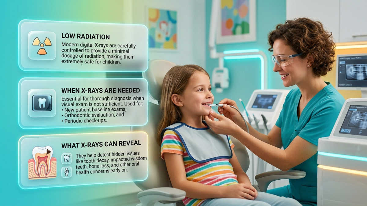
Yes, dental X-rays are considered safe for children when there is a clear diagnostic reason to take them. A single digital image delivers a very small radiation dose, usually less than a child receives from natural background radiation in an ordinary day. What matters more than the dose is the reason. The American Academy of Pediatric Dentistry states that timing should be based on each child's individual circumstances, not on age or a routine schedule.
Key Takeaways
- X-rays are diagnostic tools, not screening tools. Your child's dentist should be able to name the question an image will answer before taking it.
- The usual trigger for a first X-ray is not age. It is back teeth that have grown close enough to touch, which hides the surfaces between them from view.
- One digital bitewing is roughly comparable to less than a day of ordinary background radiation. A cross-country flight delivers several days' worth.
- National guidance changed in 2024. Lead aprons and thyroid collars are no longer recommended as routine practice, because beam collimation and digital sensors do that job better.
- You can ask questions, and you can say no. The useful question is what the image would change about your child's care, and what stays unknown without it.
Most parents nod along in the chair and then think about it later that evening. The recommendation sounded reasonable. Your child's teeth look fine to you. So why does anyone need to take pictures of the inside of a five-year-old's mouth?
Radiation is a fair thing to ask about, and the honest answer involves numbers and reasoning rather than reassurance. This guide covers what an image shows that an exam cannot, how much radiation is actually involved, how often images are genuinely needed, what your child experiences, and what to do if the visit does not go smoothly.
Why Your Child May Need a Dental X-Ray
What an Exam Can and Cannot See
A visual exam is genuinely powerful. A pediatric dentist can assess the chewing surfaces, the outer and inner faces of each tooth, the gum line, visible chips and cracks, staining, plaque patterns, and which teeth have erupted so far.
Several things stay hidden no matter how careful the exam is:
- The surfaces where two teeth touch. Once neighboring molars make contact, there is no gap for light, a mirror, or a probe tip to pass through.
- The inside of the tooth. Decay that has moved past the enamel into the softer dentin leaves little trace on the surface.
- The root and the bone around it. Infection at a root tip, a cyst, or bone changes sit entirely below the gum.
- Permanent teeth still forming in the jaw. Their number, position, and direction of travel are invisible until they erupt.
- Damage under a baby tooth. An injury can affect the permanent tooth developing directly above or below it.
An image is not a second opinion on what the dentist already saw. It answers a different set of questions entirely.
Hidden Cavities Between Touching Teeth
If your child gets a recommendation for imaging, this is the most likely reason behind it. Baby molars start out with visible spaces between them in most children. As the jaw grows, those teeth settle into contact, and the surfaces facing each other can no longer be examined visually or with a probe.
Location matters here for a second reason. Enamel on a baby tooth is thinner than on a permanent tooth, and the pulp chamber holding the nerve sits closer to the surface. Decay between two baby molars has less material to travel through before it reaches sensitive tissue, and early decay usually does not hurt. So a comfortable child is not evidence that nothing is happening.
Development, Unerupted Teeth, and Dental Injuries
The second broad reason involves development rather than decay. Between roughly ages six and twelve, your child has a mix of baby and permanent teeth, plus a queue of permanent teeth still forming inside the jaw. The AAPD treats monitoring growth and development as a purpose for imaging that is separate from finding cavities.
An image can show whether a permanent tooth is missing, whether an extra tooth is present, whether one is heading in the wrong direction, or whether there is room for what is coming. Upper canines are a common example. If one cannot be felt through the gum by around ages ten to eleven, imaging is generally how its position gets confirmed. Finding this early does not guarantee a simpler outcome, but a tooth that is starting to drift is easier to influence than one that is already stuck.
Injuries are the third reason, and one of the most frequent in practice. A tooth can look almost normal after a knock and still have a fractured root, a displaced position, or damage to the bone holding it. In younger children, a blow to a baby tooth can also affect the permanent tooth developing behind it. Here, imaging is not really optional, because treatment decisions for injured teeth depend on what happened below the surface, and timing can affect how an injured tooth is managed.
After a mouth injury
- Seek emergency medical care first if there is a possible head injury, confusion or drowsiness, difficulty breathing, or bleeding you cannot control. Dental treatment follows once your child is stable.
- Otherwise, call the dental office promptly. Note when it happened and bring any recovered tooth fragment. Dr. Leslie Jacobs Pediatric Dentistry lists 24/7 availability for dental emergencies.
Types of Dental X-Rays Used for Children
There is no single thing called a dental X-ray. Knowing which type has been recommended tells you a lot about why.
The small intraoral images at the top of that list are the workhorses, and the AAPD confirms intraoral imaging as the standard diagnostic tool for children. Two wider options exist but carry higher doses. A bitewing-style view taken from outside the mouth runs roughly three to eleven times the dose of an intraoral bitewing, so it suits specific situations rather than routine use. Cone beam CT, which produces a three-dimensional scan, is reserved for problems such as complicated trauma, impacted teeth, or surgical planning.
Digital Dental X-Rays
If your own childhood memories involve a stiff cardboard packet and a wait while something got developed, the equipment has changed. Digital sensors and phosphor plates need less radiation than film to produce a usable image, and the picture appears within seconds. That immediacy matters as much as the dose, because a poorly positioned image can be spotted and corrected rather than discovered after processing.
Digital files also transfer easily to another office or specialist, which can spare your child a repeat image later. That point comes up again below, because it is one of the few levers parents control directly.
When and How Often Children Need X-Rays
When the First X-Ray May Be Needed
You may have read that children start having cavity-detecting images around age three. That figure describes when many children reach the relevant stage. It is not the recommendation.
The AAPD is direct about this. Timing of the first image should be based on each child's individual circumstances rather than age, and taking images to look for disease before a clinical examination has happened is not appropriate practice. The exam comes first and determines whether imaging is warranted.
The practical trigger is whether the surfaces between the back teeth can still be examined visually or with a probe. While gaps remain, a dentist can inspect them directly and an image adds little. Once the teeth make contact, that access disappears. Guidance from the ADA and FDA states plainly that a child with baby teeth, no signs of disease, and open contacts may not need imaging at that visit.
This is why two children of the same age can get different recommendations on the same afternoon, and why a dentist may say your child does not need images this year but might next year. It is also a concrete thing to ask about. If your child's back teeth are still spaced, ask whether the contacts have closed yet.
Younger children are imaged less often for related reasons. They have fewer teeth, more open contacts, and a harder time holding a sensor still, and an image that cannot be interpreted delivers a dose without delivering information.
Cavity Risk and Recommended Intervals
An annual bitewing check is a common pattern, and for many children it is sensible. It sits in the middle of a published range rather than being a standard in itself. The ADA and FDA recommendations endorsed by the AAPD set intervals that depend on both dental stage and cavity risk, and they apply only when the surfaces between the back teeth cannot be examined directly.
These ranges are guidance, not automatic schedules. Higher-risk children may be imaged more often, and lower-risk children can reasonably go longer. Risk is a working assessment rather than a judgment about your parenting, and it shifts over time. Previous cavities are the strongest single predictor, alongside factors such as existing fillings, early enamel changes, deep tooth grooves, plaque levels, how often sugary or acidic food and drink appear through the day, fluoride exposure, medical conditions or medications that reduce saliva, and orthodontic appliances that trap plaque.
Worth knowing: the AAPD states that age is not on its own an indication for imaging, and says the same about insurance coverage, warning that using reimbursement as a rationale can lead to overuse. A recommendation should trace back to something specific about your child.
Panoramic Imaging During Development
Two wider images tend to come up during childhood, usually described by age because ages are easy to remember. The logic underneath is developmental.
The first arrives once permanent teeth start appearing, which for most children is somewhere around six. A panoramic view at that stage can show how the jaws are growing, whether permanent teeth are missing or duplicated, whether anything is positioned oddly, and whether there are changes in the bone. The second arrives in the mid-teens, commonly around sixteen, when wisdom teeth have developed enough for their position and available space to be assessed.
Both remain matters of clinical judgment tied to your child's development rather than automatic birthday events. A child whose permanent teeth arrive late may reach the first point at seven or eight.
How Safe Are Dental X-Rays for Children?
Here is the honest version. Dental X-rays use ionizing radiation, and the doses involved are extremely small, small enough to sit below the level at which effects can be measured in populations. Nobody responsible calls them zero risk, and nobody responsible calls them dangerous. What the profession does instead is insist that each image earn its place.
Radiation in Everyday Terms
The useful comparison is background radiation, which everyone absorbs continuously from soil, rock, building materials, food, and cosmic rays.
For the figures behind that table, a person in the United States receives roughly 3.1 millisieverts a year from natural sources, about 8 microsieverts a day, while published measurements put a digital intraoral image in children well under 5 microsieverts. The International Atomic Energy Agency summarizes it the same way. None of that makes the dose irrelevant for a child, because children are more radiosensitive than adults and have more years ahead in which exposure accumulates. That is exactly why the number of images matters more than the safety of any single one.
Lead Aprons and Thyroid Collars
This part surprises most parents, because the guidance changed recently and it changed in the opposite direction to what people expect.
In February 2024, an expert panel convened by the ADA Council on Scientific Affairs, supported by FDA medical physicists, recommended discontinuing routine use of lead aprons and thyroid collars for dental imaging, for all patients regardless of age. The AAPD's current best practice states that evidence suggests routine patient shielding is no longer necessary. The reasoning:
- The beam is now restricted to the area being imaged, so there is very little stray radiation for an apron to intercept.
- Shields do not stop internal scatter, which is where the small dose to other tissues actually comes from.
- Misplaced shields cause retakes. A collar sitting in the beam path can obscure the image, and a repeat image means a second dose.
You may still see an apron in some offices. State radiation regulations vary and are being reviewed, and some practices continue the habit because parents find it reassuring. Neither approach means your child is being poorly cared for. If you notice a difference from what another office did, it is fine to ask what protocol is followed and why.
How Dentists Minimize Unnecessary Exposure
Dose reduction is a package rather than a single upgrade. Faster digital receptors, restricting the beam to the size of the sensor, positioning devices that reduce unusable images, and limiting the number of images to what is diagnostically necessary all work together. The combined effect is that a modern set of bitewings represents a fraction of what the same set involved decades ago.
Which leaves the decision itself as the biggest remaining lever. Professional guidance frames imaging as a way to substantiate or rule out something the clinician already has reason to suspect. In practice, that means categories such as pain or trauma, suspected or visible decay, large or deep existing fillings being monitored, eruption and development concerns, swelling, and unexplained symptoms like sensitivity or bleeding. The AAPD also asks clinicians to weigh the realistic likelihood of obtaining a usable image from this particular child today, which is why a dentist may choose to wait rather than attempt one.
Using Previous X-Rays
This is one of the few ways to reduce your child's lifetime exposure that sits entirely in your hands. The AAPD states that reviewing prior images, including those obtained through records transfer, contributes to the decision about whether new imaging is necessary.
If your child has been imaged anywhere in the past couple of years, including at a previous dentist, an orthodontic consultation, or an emergency visit, mention it when you book rather than when you arrive. Ask the previous office to send the actual image files with their dates. Practices cannot always use an outside image, since quality varies and the angle may not show what is needed, but sometimes it works and asking costs nothing.
What a Dental X-Ray Appointment Is Like
What Your Child Experiences
Pediatric practices generally use an approach called tell, show, do. Your child is told what will happen in words matched to their age, shown the equipment, allowed to touch the sensor or bite tab, and only then asked to try it. The unfamiliar object becomes a known object before it goes near their mouth. The language is chosen deliberately too, which is why a sensor might be described as a camera that takes pictures of teeth.
For a bitewing, your child sits upright, a small sensor on a tab is placed beside the back teeth, and they bite gently to hold it in place. The tube head is positioned outside the cheek, the team member steps behind a barrier, and the exposure lasts a fraction of a second. For a panoramic, your child stands or sits at the machine, bites on a positioning guide, and stays still while the arm travels around their head. Some children find that easier because nothing goes inside the mouth. Others find standing still harder.
The most useful thing you can do at home is keep your framing neutral. Telling your child there is nothing to be scared of introduces the idea of being scared. Saying the dentist is going to take a picture of their teeth does not.
Does It Hurt?
No needles and no drilling are involved, and there is no sensation from the radiation itself. The honest caveat is about the sensor rather than the X-ray. A rigid sensor has edges, and in a small mouth those edges can press against the gum, the floor of the mouth, or the palate. Some children find it merely odd, some find it uncomfortable for the few seconds it is in place, and a few gag.
Telling your child it will not feel like anything sets them up to distrust the next thing they are told. Telling them it might feel hard and strange but finishing quickly holds up better. Also tell them they can say so if it bothers them, because knowing they have a way to stop is often what makes a child willing to start.
How Long It Takes
Positioning takes longer than exposure. A set of four bitewings usually occupies a few minutes from start to finish, and a panoramic image takes a minute or two, including getting your child lined up. Within a routine preventive visit that also includes the exam and cleaning, the imaging is a small fraction of the total.
If You Are Pregnant
You are not the one being imaged, so the question is not about shielding you. It is about where you stand. Standard radiation safety practice is that anyone whose presence is not required steps behind the protective barrier during the exposure, which is why the operator does the same. Following that means your exposure is essentially nil.
Tell the team when you arrive, so nobody asks you to hold a sensor or steady your child's head. If your child cannot manage without a hand to hold, ask whether a holding device or another adult can do the job.
What If Your Child Has Trouble With X-Rays?
Some children manage imaging on the first try, and some do not. A child who cannot tolerate a sensor is not misbehaving. They are having an ordinary reaction to something unfamiliar in a sensitive part of their body, and pediatric practices plan for it.
Gagging, Anxiety, and Movement
Gagging is a reflex rather than a choice, and some children have a trigger point much further forward than others. Trying harder makes it worse, because the child braces before the sensor is even placed. What tends to help:
- A smaller sensor. Child-sized receptors make a substantial difference in a small mouth.
- Sitting upright rather than reclined. Being tipped back tends to intensify the reflex.
- Steady nose breathing during placement, which is one of the more reliable techniques.
- One image at a time, with a practice run first while the machine is switched off.
Fear and restlessness are handled with communication rather than force. Tell, show, do, positive reinforcement for small steps, distraction, and desensitization across more than one visit are the usual tools. A child who cannot manage a sensor today may manage it easily three months later. Splitting the work also helps, since two images today and two next time leaves your child with a memory of having succeeded rather than failed.
One point deserves stating clearly. Sedation is not a routine answer for imaging on its own. Sedation decisions involve individual assessment of medical history, airway, development, and the actual treatment needed, weighed against real risks. It may enter the conversation when imaging is part of a larger treatment plan, but it is not a solution to a difficult sensor placement.
Sensory or Special Healthcare Needs
For a child with sensory sensitivities, a developmental difference, or a complex medical history, the sticking point may be bright lights, the texture of a sensor, being asked to stay still, or an unpredictable sequence of events. There is no single accommodation that works, because a diagnosis does not determine what a particular child finds difficult. Planning individually is what works.
- Call ahead and describe what specifically tends to be hard, rather than only naming a diagnosis.
- Ask about a quieter appointment slot, and about visiting the room and handling a sensor before anything is attempted.
- Bring headphones, sunglasses, or whatever your child uses to regulate, and ask whether they can stay on.
- Say plainly what has failed before and what has worked. It saves everyone a repeat of the failure.
Dr. Jacobs is extensively trained in the care of children with special needs, and the team includes staff fluent in English, Spanish, and sign language. If communication access matters for your child, mention it when you book so it can be arranged.
What Happens If an Image Cannot Be Taken
The AAPD acknowledges that a radiograph can be clinically indicated while a diagnostic image cannot actually be obtained. When that happens, the expectation is that the dentist explains the limitation to you, documents it, and decides based on how urgent the treatment need is and the relative risks of proceeding without the image.
In practice, that usually means treating what is visible and trying again next visit, or arranging a short appointment focused only on imaging. Either way you should leave knowing what remains unknown and what would prompt a change in plan. A visit that ends without the image is not a wasted visit, because your child learned that nobody forced anything.
Understanding the Results
Reading Light and Dark on the Image
An X-ray image is a map of density. Dense material absorbs more radiation and appears bright or white, while less dense material lets more through and appears dark. Enamel and any metal are the brightest things on your child's image; bone reads as textured mid-grey, and the pulp chamber inside the tooth shows as a darker channel.
Decay matters here because it softens tooth structure and reduces density. That is why a cavity between two teeth often appears as a dark notch cutting into the bright band of enamel where the teeth touch.
Common Findings
- Decay between teeth, the most frequent finding and the usual reason for the image.
- Decay beneath an existing filling, sometimes visible as a shadow under an earlier restoration.
- A dark area at a root tip, which can indicate infection spreading into the surrounding bone.
- Roots of a baby tooth dissolving, which is normal development as the permanent tooth beneath pushes up.
- An unerupted, missing, or extra permanent tooth, or one angled in a way that may cause trouble.
- Crowding or space concerns, including wisdom tooth position in the mid-teens.
Something appearing on an image does not automatically mean something must be done to it today. Several of these are monitored rather than treated, and one of them is simply development proceeding normally.
What Happens Next
If decay is found, the response is not one-size-fits-all. Recommendations depend on how deep the lesion is, which tooth it is in, how long that tooth is expected to stay in the mouth, whether the nerve appears involved, and your child's overall risk picture.
Shallow early changes are sometimes managed by strengthening the enamel and watching rather than by drilling. Established decay in a tooth that will be in place for years usually needs restoring. A baby tooth close to falling out on its own may be handled differently from the same tooth at the same depth three years earlier. Restorative options for children vary considerably, and choosing among them is a conversation rather than a verdict.
Questions Parents Can Ask
Bring these to the screen while the images are up. Each takes about a minute to answer.
- Can you show me on the image what you are describing?
- What did this image reveal that the exam could not?
- Does this need treatment now, or monitoring?
- What are the alternatives, including waiting?
- When would you expect the next images, and what would make that sooner or later?
It is also worth asking for a copy of the images for your own records. That makes any future transfer easier and can spare your child a repeat image years from now.
Can You Decline an X-Ray?
Yes. This is your decision, and asking about it does not make you an obstructive parent. Informed consent means being told what is proposed, why, what the alternatives are, and what happens if you decline. Declining is one of the available answers.
You can ask why now, ask whether fewer images would answer the question, and ask to think about it and call back. You can also change your mind in either direction, because circumstances change and so do teeth. A good practice will tell you plainly what the limitation means for your child's care, note the conversation, and carry on caring for your child.
What Can and Cannot Be Diagnosed Without One
This is where the real trade-off sits, so it is worth being clear-eyed about it.
Without imaging, a dentist can still assess visible decay on open surfaces, gum health, plaque and calculus, visible chips and cracks, which teeth have erupted, bite relationships, and obvious swelling. For a child with spaced teeth and no symptoms, that may cover nearly everything relevant.
What cannot be reliably assessed is decay between teeth that are in contact, how deep an existing lesion has traveled, whether infection is present at a root tip, the condition of teeth still forming in the jaw, whether a permanent tooth is missing or extra, and injury below the gum line after trauma.
So declining does mean nothing can be done. It means accepting diagnostic uncertainty in those specific areas, and accepting that anything developing there will likely be found later and be larger. For a low-risk child with open contacts, that is a small trade-off. For a child with several previous cavities and closed contacts, or one who has just taken a hit to the mouth, it is a bigger one. That difference is the thing worth discussing, and it is easier to discuss on the phone before the visit than in the chair with your child listening.
Cost and Insurance for Children's Dental X-Rays
Prices vary by region, practice, and what is included, so no article can quote a figure that will be accurate for your family. What drives the number is straightforward: how many images are taken, which type they are, whether they are bundled into a preventive visit or itemized separately, and whether existing images can be transferred instead of repeated. Panoramic images are generally billed separately from intraoral ones, and three-dimensional imaging costs considerably more than either.
Dental plans usually treat imaging as diagnostic care and often cover it at a higher rate than restorative work, subject to frequency limitations. Many will pay for bitewings only once per year or once every two years, and for a panoramic image only once every several years.
Those limits are contractual terms, not clinical recommendations. A plan that covers bitewings annually is not saying your child needs them annually, and one that covers them every two years is not saying your child cannot need them sooner. The AAPD states directly that insurance coverage is not on its own an indication for imaging, and warns that using reimbursement as a rationale can lead to overuse. Before the appointment, it is worth confirming with the office whether they are in network, what your plan's frequency limit is, and whether your child has already used that allowance elsewhere this period.
Frequently Asked Questions About Kids' Dental X-Rays
Are Dental X-Rays Safe for a Toddler or a Baby?
Imaging is used in very young children, but rarely and for specific reasons such as an injury, visible decay, or a tooth erupting oddly. The dose is very small at any age. Toddlers are imaged less often because there is usually less that cannot be examined directly, not because the dose is unsafe for them.
Can I Stay in the Room With My Child?
In many pediatric offices, yes, though anyone not needed steps behind the barrier during the brief exposure itself. Policies vary, so ask when you book. Some children do better with a parent visible, and some do noticeably worse, and if the team suggests waiting just outside, that suggestion is about your child rather than about you.
How Many X-Rays Is Too Many in One Year?
There is no published number, because it depends on what is happening in your child's mouth. A more useful test than counting is whether each image had a stated purpose. The intervals in the table above give you a reference point to compare against.
Do Baby Teeth Show Up on an X-Ray the Same Way Permanent Teeth Do?
They appear the same way, since both absorb radiation according to density. What differs is the reading. Baby teeth have thinner enamel and a pulp chamber closer to the surface, so decay of the same depth is proportionally more significant. Their roots also dissolve gradually as the permanent tooth moves up, which is normal rather than damage.
What If My Child Had X-Rays at Another Dental Office?
Say so when you book and ask the previous office to send the image files with their dates. Existing images sometimes make new ones unnecessary. They cannot always be used, since the angle or quality may not answer the current question, but the request is free.
Can an X-Ray Show Whether My Child Will Need Braces?
It contributes pieces of the picture rather than answering the question. A panoramic view can show whether permanent teeth are all present, whether any are extra or badly angled, and how much room exists. Orthodontic assessment also depends on how the teeth meet, jaw function, and facial growth, none of which an image shows.
Pediatric Dental X-Rays in Lafayette, LA
The dose from a digital dental image is genuinely small. The question that deserves your attention is not whether the dose is safe, but whether this particular image, for your child, right now, will answer a question that matters. When a recommendation traces back to something specific, such as closed contacts, a fall last weekend, or a permanent tooth that has not appeared, it is doing exactly what professional guidance intends. When it cannot, you are entitled to ask what prompted it.
That judgment can only be made after an examination, which is why Dr. Leslie Jacobs Pediatric Dentistry assesses each child individually before recommending dental imaging. Dr. Leslie Jacobs is a board-certified pediatric dentist serving families in Lafayette and the surrounding Acadiana area.
You do not need to wait until your child is in the chair to ask about imaging. Raise it when you book. Mention any images taken elsewhere so records can be requested, and tell the team what tends to be difficult for your child. To arrange a visit, contact the practice or call the Lafayette office.
About this guide
- This is general information for parents, not a diagnosis or treatment recommendation for any individual child. Whether your child needs imaging can only be determined after a clinical examination.
- Clinical points follow the AAPD best practice on prescribing dental radiographs, the ADA and FDA recommendations it endorses, and the 2024 ADA recommendations on radiation safety. State radiation regulations vary, so protocols at individual offices may differ.
.jpg)
.jpg)


